The Effects of Light Pollution on the Environment

by: Kahsennaró:roks Deom

What is Light Pollution?

Light pollution is defined as the artificial, man-made light that ‘contaminates’ the purity of the night sky.1 At first, this may seem like a non-issue, but it doesn’t just prevent everyday civilians from looking at the stars at night. Light pollution also means that it impedes on the health of humans, mammals, insects, and even plants – directly or indirectly (Not to mention it probably affects astronomers and other scientists – but that is another topic). The question “what is light pollution?” isn’t simply answered by defining it. As a community, those who are interested in the topic must understand how light and its pollution is measured, who it is affecting, how it is affecting them, and how to control this issue. Hopefully this post will help those interested in light pollution learn more on how they can combat the issue.

How to Quantify Light Pollution?1

To start, you must know that light pollution isn’t a problem all the time. It is only considered a problem when it is night time (when the sky is dark), and it depends on the amount of natural light that is available. So, an unpolluted area is an environment illuminated by natural light only, very little artificial light of a certain wavelength, or not illuminated at all. This area is usually outdoors (or indoors when light is not compromising health). A polluted area can be indoors and outdoors, and is highly illuminated by artificial light of a short wavelength.

The addition of artificial light is measured by concentration in volume, emissions, direction, spectrum, relativity, and physiology of vision. Some of these measurements will be discussed further in the text.

As mentioned above, short wavelengths (blue and green) are more severe for both viewing the night sky and the health of the planet. This is because short wavelengths indicate time of day for certain organisms while also affecting metabolism and other functions. They also affects our view of the night sky because they produce the most sky glow (diffusion of photons by moisture and air particles, resulting in a brighter sky).

Who is Affected?

Humans bear significant consequences of light pollution, but we aren’t the only ones. Along with use, mammals, birds, insects, plants, and many more are affected by artificial light at night

Humans are affected by light pollution because it impacts melatonin production, which impedes on our natural sleep cycle and has been linked to higher rates of breast cancer and prostate cancer. Our human biology has depended on the light/dark cycle adaptations, as all organisms’ biology is. In summary, our ancestors were active during the day, with the help of natural light, and restored their energy at night, with the help of the dark. Our bodies are adjusted to this cycle. One process that humans rely on that is aided by the day/night cycle, is pineal melatonin. This hormone is created in the pineal gland (the brain) and distributed to various parts of the body that use it. For instance, we use melatonin for sleep – it decreases vigilance and increases fatigue. It also repairs DNA, and manages or pituitary and ovarian hormones. These repairs happen when we sleep.

Artificial light was popularly introduced not too long ago (around 120 years). With this fast development, our biology hasn’t been able to adapt as quickly as it did to the natural light cycle. Due to the introduction of artificial light seeping through our windows at night, LED phone screens, etc., melatonin production is being suppressed. This causes our night/day cycles to lag (why one might be tired during the day even after a good night sleep), reduces fatigue, and reduces the amount of melatonin produced. this means that the areas in our bodies that need repair, aren’t being attended to at the same rate.

According to Haim et al. (2013), in a study, mice were treated with male prostate cells and were put under different light conditions. Some mice with shorter artificial light days were given another 30 minutes of light after being in the dark for 7 hours. Some mice with longer days of artificial light were treated with melatonin during the dark period. Those with the longer days had larger tumors than the short-day mice, and they also had lower melatonin levels. this shows that a longer exposure of artificial light increases the risk of cancer.

Other studies showed hat women with lower levels of melatonin were had larger tumors than women who were otherwise healthy, and were more at risk of cancer than others.

Nocturnal Predators, Migratory Birds, and food webs

At night, in terms of short term safety, artificial light is great for humans for visual aid; however; it shouldn’t be shocking that other animals would disagree!

Nocturnal mammals3:

  • They have a high amount of rods in their eye biology, allowing for high visual stimulation by a low number of photons.
  • Having few cones means they are easily blinded by bright lights.
  • Dark nights are ideal for activities such as hunting.
  • On bright nights, prey stay in the out of the moon light, for there are other predators that are not necessarily nocturnal
  • With increased artificial light impeding on wildlife habitats, nocturnal predators are less likely to see their surroundings and are unable to catch as much prey, due to the prey’s preferred area of safety.
  • Nocturnal animals don’t hunt in complete dark, so hunting with brighter light (depending on wavelength and intensity) can lead to higher accuracy, but less prey would be caught.

Migratory Birds3:

  • It has been noticed especially by lighthouse keepers that birds tend to be attracted to light.
  • Hunters used to use light to attract birds, and birdwatchers use light now to grab the attention of birds.
  • Some songbirds travel at night to avoid the presence of predators.
  • Birds are attracted to highway lights, city lights, radio towers and airplane towers.
  • This attraction leads many birds to collide with buildings and structures, killing millions each year5.

Pea aphids and Great bird’s foot trefoil4

  • The pea aphid’s food source is a flowering plant called the “Great bird’s foot trefoil”.
  • In a study done at the University of Exeter in the United Kingdom recorded the effects of artificial light on the food web between the aphid’s predator, the carabid beetles, aphids, and the trefoils
  • With and without the presence of the aphid, the trefoil density decreased due to both white light (LED) and amber light (HPS)
  • Aphid presence also decreased due to both lights.
  • There was no recorded changes in the predator’s presence, but in another observation noted in this study, the carabid activity increased, which had consequences on its prey and in turn, on the prey’s food source.

Costa Rica: Turtle-Hatching 6

In Costa Rica, we had an opportunity to observe sea turtles hatching their eggs at night by the ocean. We learned there that when the turtle eggs hatch, the hatchlings wait until dark to make their way into the ocean. This is because they depend on the moon’s reflection on the water – they look for the brightest horizon to find the ocean. When there are other light sources near the beaches, such as city lights or lamp posts for beach visitors, they can mistake that source as the brightest horizon. With excess light, they are exposed to predators, and can lose themselves by losing track of the ocean.

Different Light Types7,8

As mentioned above, there are many different light types – different wavelengths showing different colours. Some lights produce colors of short wavelengths, while other have longer wavelengths. Shorter wavelength-lights, such as LEDs, are shown to have worse effects on vision and biology compared to longer wavelength-light, like LPS (low pressure sodium).

Different light types range from LEDs, PCA LED (Phospho-converted amber LED), NBA LED (narrow band amber LED), HPS (High pressure sodium), and LPS.

What is Being Done?

Flagstaff, Arizona9:

  • Started the Dark Skies Coalition – spread awareness of light pollution and promote dark nights.
  • The city has installed lights that are covered on the top, as to not travel upwards and impede on view.
  • Using mostly amber lights, with high wavelengths.

Natural Sounds and Night Skies division of the National Parks10:

  • Also promoting dark night skies – not only for visitors at the National Parks, but also for those who live in national parks (AKA not humans)
  • Reduce amount of light used in parks (mainly only on the paths for visitors).
  • Lights are shielded and there is no blue light.
  • Yellowstone National Park also educates its visitors on astronomy and the increasing problem with light pollution.

Sources:

1 Hollan, Jan. “What is Light Pollution and How do we Quantify it?”. N. Copernicus Observatory and Planitarium. Dec 2006.

2 Haim, Abraham, Boris A. Portnov. “Light Pollution as a New Risk Factor for Human Breast Cancer and Prostate Cancers”. Springer. 2013.

3 Rich, Catherine, Travis Longcore. Ecological Consequences of Artificial Night Lighting. Island Press.

4 Jonathan Bennie, Thomas W. Davies, David Cruse, Richard Inger, Kevin J. Gaston. Cascading effects of artificial light at night: resource-mediated control of herbivores in a grassland ecosystem. Philosophical Transactions of the Royal Society B, March 2015.

5 Ebersole, Rene. “Collateral Damage”. Audubon. Jul-Aug 2014.

6 “Sea Turtle Conservation”. International Dark Sky Association.

7 “LED Lighting and Dark Skies”. Flagstaff Dark Skies Coallition. 24 Apr. 2018.

8 “The Color of Lights: More than Meets the Eye”. Illinois Coalition for Responsible Outdoor Lighting. Jan. 2011.

9 “Home”. Flagstaff Dark Skies Coallition. 2 May. 2018.

10 “Night Sky Initiatives: Yellowstone National Park”. National Park Services.

11 Howell, Elizabeth. “‘Sky Glow’ Kickstarter Takes on Light Pollution of the Night Sky”. Space. 8 May. 2015.

12 “Winding Through Highway 1 and Redwood Forest at Night”. Youtube, JesdaG, 25 May. 2018.

13 Rich, Catherine, Travis Longcore. Ecological Consequences of Artificial Night Lighting. Pg. 79, Island Press.

14 “Lamp Spectrum and Light Pollution”. Flagstaff Dark Skies Coallition. 2 May. 2018.

Prevention Method of Spreading Ophryocystis elektro-scirrha Spores in Laboratory Reared Monarch (Danaus plexippus) Population

Maggie Blondeau, Nicholas Danopoulos, Alexandre Pham, Suthan Sinnathurai

Abstract

1) Because of the 78.35% mortality rate of monarch butterflies in the 2016 Monarch Project at Dawson College, a solution to reduce the cross contamination of the sporing parasite (Ophryocystis elektroscirrha; OE) causing the deaths was found. We believe washing hands with common soap will reduce the number of OE spores found on human skin.

2) OE is a protozoan parasite that as a spore on the wings and body of a grown butterfly, is introduced to milkweed plants when the monarch lands on it[2]. OE either kills monarch pupas during metamorphosis, or the monarch emerges with other OE induced consequences (e.g. disfigurement).

3) 3 trials of washing hands after handling OE infected monarchs to reduce the amount of spores attached to the skin were performed.

4) The average amount of OE spores present on fingers before hand wash and after hand wash were 488.3 and 0.66 spores per millimetre squared respectively.

5) There is a significant difference between the amount of spores per millimetre squared before washing hands versus after. This shows how using common hand soap can reduce spores found on fingers by 99.9986%, helping to reduce the cross contamination of OE spores due to human contact.

Introduction

         The monarch butterfly (Danaus plexippus) is a species in danger of extinction, with one study showing that there was an 84% decrease in the Eastern North American monarch population between 1996 & 2015. The decline in the population is thought to be due to the use of insecticides, loss of breeding habitat, plants, climate change, and decrease in the amount of milkweed (genus Asclepias)[1]. Humans are the cause for several of these factors, and therefore have the responsibility to help revive the number of butterflies in its population. The 2016 Monarch Project at Dawson College had a 78.35% mortality rate of monarch butterflies, believed to be caused by a parasite called Ophryocystis elektroscirrha (OE). Previous years also experience increased mortality rates, which may have also been caused by this parasite. With such a large mortality rate, the community needs a safer method of handling the monarchs and their environment, due to the easily spreadable nature of OE spores (Figure 1). If a method of diminishing spores is found, it will decrease the number of deaths due to this parasite for future years of this project.

Ophryocystis elektroscirrha (OE)

         OE is a protozoan parasite, that as a spore on the wings and body of the grown butterfly, is introduced to milkweed plants when the monarch lands on it. These spores may also cover monarch eggs if they come in contact with an infected butterfly. Spores are then consumed by the monarch caterpillars as they feed on the milkweed. Once ingested, they release sporozoite (the cells which infect the pupa during metamorphosis) as the larva produces digestive chemicals to break down the milkweed. While the caterpillar is in metamorphosis, these cells go through the stages of vegetative schizogony (asexual reproduction) and line the intestinal tract of the pupa. The parasite continues to reproduce until several days before the butterfly emerges from its cocoon, where it then begins to produce spores[2]. Once infected with this parasite, there are three possible outcomes: The pupa dies in its chrysalis, it emerges as a disabled/disfigured butterfly, or the butterfly emerges without any physical irregularities, though it carries the spores to the next milkweed plant it lands on, allowing the cycle to restart[3].

How to Prevent Humans from Spreading Spores

         Monarchs may not be the only species which contribute to the spread of OE spores. It is possible for the spores to be carried from one plant to another, or one butterfly to another, due to human cross contamination. Spores attach to human skin, as studies and personal observation suggests[4], therefore if a person works with a milkweed plant, or a butterfly/caterpillar contaminated with OE, there is a potential for the spores to be spread to any other plant or butterfly/caterpillar that the person may work with. This project aims to find a way to prevent, or diminish this cross contamination, as it contributes to the mortality rate of monarch pupae and to the population of disfigured monarch butterflies.     

         There have been experiments performed to test hand washing to reduce the amount of spores found on a person’s skin, however OE was not the spore in question. One experiment was performed on Clostridium difficile, a sporing organism that can be spread through human cross contamination[5]. One study found that a non-antimicrobial hand wash reduced the amount of spores found on a carriers hands by 78%[6]. This experiment is pertinent, as it proves how a basic store bought hand soap can eliminate the majority of spores found on human hands. Therefore, it may reflect how the experiment of this project will perform, and thus will reduce the cross contamination of OE spores by humans.

         The project aims to determine whether the use of store bought hand wash (a common type found in homes and laboratories) will reduce the number of spores on the person’s skin, which in turn would limit the spreading of these spores. This research has never been done before to our knowledge, and therefore the results will help inform everyone working with monarchs and/or milkweed plants. Taking steps towards reducing human cross contamination of OE will help the struggling monarch population. Therefore, this project will help and improve the survival rate of butterflies of the Monarch Project at Dawson College.

Methods

         Before the experiment, the participants’ hands were washed (SoftsoapTM) and dried completely. The participants then rubbed the OE infected monarch’s abdomen with their index finger and thumb. Afterwards, one piece of scotch tape was accurately applied to the part of the index finger that came in contact with the monarch. It was then pulled off and stuck to a hemocytometer slide identified that it is the slide used before the final hand washing. Their hands were then washed and dried again. A different piece of scotch tape was applied to the thumb that came in contact with the monarch. The piece of scotch tape was stuck to the hemocytometer slide and identified as the slide used after the final hand washing. There were three participants, and therefore three trials. Error bar analysis[7] was then used to determine whether the two groups are significantly different.

Results

         The average amount of spores present on fingers before final hand wash and after final hand wash were 488.3 and 0.66 spores per millimetre squared respectively after 3 trials (Figure 2). The error bar analysis[7] used concluded that the two groups are significantly different, with a gap of 258.01 spores present.

Discussion

         The average amount of OE spores present on fingers before final hand wash and after final hand wash were 488.3 and 0.66 spores per millimetre squared respectively (Figure 2). The two groups had varied standard deviations, with the before group’s being 397.2, meaning that the average from all 3 trials were largely varied. This could have been caused by the limited material (i.e. only one OE contaminated monarch was used in all trials). Therefore the majority of the spores may have been removed during the first trial, with the following trials having fewer spores to attach to the skin. In a more preferable case, there would be a different contaminated monarch for each trial. However, the standard deviation of the after group was low, being only 0.57, suggesting that the hand wash (SoftsoapTM) did remove almost every spore present on the finger. The standard errors were also varied, with that of the before group being high, at 229.3. The after group had a standard error of only 0.33. The standard error bar analysis[7] used concluded that the two groups are significantly different, with a gap of 258.01.

         The result effectiveness of the hand wash is similar to that of the experiment performed on Clostridium difficile, a sporing organism that can be spread through human cross contamination. The effectiveness of a hand wash on removing C. diff spores was 78%[5], and the percent effectiveness of the hand wash used for this experiment at removing OE spores was 99.9986%. This suggests that certain sporing organisms are removable via hand wash, which is pertinent to all those who work in environments potentially containing sporing parasites.

         This study has never previously been performed, and thus this research is an important and impactful addition to the community of individuals who work with monarchs and/or milkweed by preventing the cross contamination of OE spores. The question remains of which components in the hand soap allow for the decrease of spores attached to the oils human skin secretes, as well as if any hand soap will do this, or if specific brands are required.

References

1. Schmitz, B. A. (2016). Pollinator Populations may become Extinct. Wild Ones Journal, 29(3), 16.

2. Sander, S. E., Altizer, S., De Roode, J. C., & Davis, A. K. (2013, August 28). Genetic Factors and Host Traits Predict Spore Morphology for a Butterfly Pathogen. doi:10.3390/insects4030447

3. Altizer, S. M., & Oberhause, K. S. (1998, August 10). Effects of the Protozoan Parasite Ophryocystis elektroscirrha on the Fitness of Monarch Butterflies (Danaus plexippus). Journal of Invertebrate Pathology, 76-77.

4. Smith, E. (2013, December 22). OE – Ophryocystis elektroscirrha – Monarch Butterfly. Butterfly Fun Facts.

5. Gouliouris, T., Brown, N. M., & Aliyu, S. H. (2011). Prevention and treatment of Clostridium difficile infection. Clinical Medicine, 11(1), 75-79.

6. Shrestha, N. (2009, April). C. difficile spore removal via hand washing a challenge. Infectious Disease News. p. 23.

7. Cumming, G., Fiddler, F., & Vaux, D. L. (2007, April 9). Error bars in experimental biology. The Journal of Cell Biology. doi:10.1083

Case Study on Mount-Royal park

How to protect our urban ecoterritory

By Loubna Hiba Labdeli, Kuang Jin

Human often forget they are a part of nature. In fact, many have this mentality, that human is at the opposite side of nature. In our history books, nature is often portrayed as a monstrous being, a treat for early human. Then, human by using its strength and intelligence has overcome challenges, conquers and finally dominates the natural environment. From the fire of Prometheus to the Great Flood of Gun-Yu, this pattern of origin myth has deeply rooted in our culture and result in human both fear and lack of respect toward the nature. Until today, word like “wild” or “wilderness” still have some sort of negative connotation due to that reason.

As a modern human born and raised in the cities, it’s truly hard to develop a sense of solidarity and affinity with nature due to our lack of exposure to any type of natural environment. This lack of connection with the wilderness has activate our fear for the unknown and distance ourselves even more from the nature. This might seem like a minor issue, but when comes to environmental conservation which needs the support and the collaboration of the whole society, this will have a major impact on public’s opinion. More specifically, this distance will become an element of hindrance. People generally valued the direct and the indirect value of biodiversity. But many fail to comprehend when comes to the existential value of biodiversity. This absence of empathy toward other organism find its origin in the unfamiliarity toward nature and the fear that comes with it. To balance out this fear, many modern humans have this almost delusional perspective of human capability, they think human technology could do anything, they don’t need to respect the balance in nature and could still get what they need. This ignorance born out of blind arrogance makes human act slowly in front of serious environmental crises like climate change.

This is not only an environmental problem, but also a cultural problem. Culture is essentially the sum of how people think and what people do, it is dynamic and undergo constant changes, but to drive the change in culture is difficult. In fact, the only way to shape culture is through education. To break this fear and unconsciousness toward the nature, we must build up affinity and connection between nature and human. By that, I mean to integrate and merge nature into human life.

A very straightforward way is to built parks into cities. There are many types of parks, some are nothing more than an open grassland that have very poor diversity, they are not subject of this discussion. What we are interested at is a biodiverse area with in cities that could be treated as a natural reserve. We could call these parks with the term ‘ecoterritory’. But these ecoterritory need to be managed wisely, the dynamic yet delicate balance between biodiversity and human activity need ecological knowledge and care to maintain.

Mount-Royal Park located in the center on Montreal island is one of the most iconic and successful ecoterritory in Quebec. It is not a coincidence that after almost 400 years of human disturbance, this landmark of Montreal surprisingly still maintains some degrees of biodiversity. This result is due to historical and ongoing conservation effort that been put in to this tiny mountain. Even though Mount- Royal Park still face some challenges and difficulty, but with the Montreal city development master plan, we can expect a bright future for the park. The conservation of Mount-Royal is not only beneficial for Montreal but could be a good example for the management of other urban ecoterritory. This park is a valuable place that contains an important number of native red Oaks in strands which provide a valuable breeding habitat for bird life and may constitute the home of rare plant species It is also a high visited park with 13% of its users being tourists, one could not ignore the economic aspect

of this touristic attraction. The promotion of the park’s enhancement
and protection also is in the interest of the public health as it is a place that
allows them to go hiking or go out. The Mount-Royal park represents also an important air purifier as it contains a high forest density clearing the
polluted air of the Metropol. And its high biomass helps shade the city from the sun, allows evapotranspiration and also helps reduce the temperature of the air as shown in the figure 1. Therefore it have the ability to regulate local climate. These properties has made Mount-Royal a refuge during the age of the first industrialisation, in 1861 Hôtel-Dieu de Montréal has moved from the old port to settle in the mountain to get away from the heavy pollution, then followed by Royal Victoria Hospital at 1893 and Shriners Hospital for Children at 1925. Furthermore, it has significant value on the population’s education as we said, especially the younger ones as it could be used to promote the importance of sustainable management and the encouragement in the usage of natural parks and the mountains’ valuable heritage.

This important landscape, however, hasn’t always been protected as it should. In the past centuries, almost 100% of the landmass was altered by human. In the full swing of industrial development, Montrealers has first realized the importance of preserving this important natural and cultural heritage at the second half of the 19th century, this lead to the inauguration of Mount Royal Park at 1876. This initiated the preservation of Mount-Royal’s natural environment, but it has offered only minor protection for the ecosystem due to the lack of ecological knowledge and effective regulation in management. Therefore at the middle of 20th century, Mount-Royal’s ecosystem face serious problems. The endless construction on the mountain has result habitat loss, there was a significant decline in the size of forest, the diversity of the ecosystem and overall biomass (Figure 2). The launch of the Mount-Royal park has introduced and increased human disturbances, resulting lost in the forest undergrowth that we could still observe today. Lack of ecological knowledge lead to the introduction of many invasive species like Norway maple (Acer platanoides) which was brought in for street greening, selected for its small root radius, but soon outcompete local species and treat the indigenous sugar maple population (Acer saccharum). Until recent years, the increase of international transportation has introduced many fungal diseases into the area, such as the fungal pathogen Hymenoscyphus fraxineus from Europe which has caused massive ash dieback in Mount- Royal .

To solve those issues in order to preserve the forest of Mount-Royal and keeping Montreal an ecologically functional city, conservation plan need to be designed wisely. With the Montreal city development master plan put out at 2004, city suggest that ecoterritory should be considered and managed as natural reserves within the city. 10 ecoterritories was identified specifically as independent but interconnected conservation subject according to Politique du Patrimoine Art.3.59 of Montreal. These ecoterritoires as a unified network will not only help strengthen the resistance of each ecoterritory, but also facilitate species migration. for example, the Île Bizard ecoforest corridor allow species to enter the ecoterritory network from the north, then by passing through the ecoterritory of Cheval Blanc rapids and Bertrand stream basin species could bird could enter the Mount- Royal area. After that, between the ecoterritory of mount-Royal and the reserve of Lachine des Rapides, There is the Saint-Jacques escarpment which act as a stepping stone, this opened up a north-south pathway for bird migration and potentially this pathway could benefit species in climate change caused range shift to move toward the north. (figure 3,) Usually urban areas will block migration and fragment habitat, but with these ecoterritories, a city could reduce its impact on the environment. Within Mount-Royal Park there is a similar network, directed by the Mount-Royal Protection and Enhancement Plan this natural reserve is divided in 3 main conservation area called core zone, which are connected with ecological corridors. A layer of buffer zone of 30m cover the edge of the core zone (Figure 4). In those core zones, constructions are forbidden by law, in the corridors construction is at least regulated to prevent further habitat loss. The conservation team (ami de la montagne) also attempt to reconstruct the three stratas structure of the forest in order to increase the habitat heterogeneity and maximize the biodiversity. To reconstruct the lost of undergrowth in some areas due to human disturbance, the park has recently set up re-naturalisation area in the south side of the mountain to avoid human entree. The plan has also put highlight on the integrity of indigenous species, the park remove regularly invasive species, in the last year, Mount-Royal park has cut down almost 200 individuals of Norway maple and replaced them with native sugar maple.

Even with all these conservation effort that went into the park, there are still some challenges remain, the integrated tourism has inevitably caused the presence of road crossing the core areas and that lead to fragmentation and road side effect (specifically the increase in invasive herbaceous overgrowth, decrease in indigenous undergrowth ) There might be the need in the future to construct micro-ecological corridor to increase habitat availability and to define more buffer zone at the road edge. The re-naturalisation area is also having hard time to recover its undergrowth due to the higher strata blocking the sunlight. This is because the Mount-Royal have a very special land relief pattern, the slope of the mountain is almost in a stairs like pattern, which means there are both very sharp rise and flat surface in the landscape. In forest, trees grow in colonies, tree of similar taxas and age will have similar height, many trees of same height stand on a flat surface will inevitably block the sunlight from reaching the lower stratas. This type of relief might not be beneficial for plant undergrowth, but it is coincidentally appropriate for the construction of artificial wetlands, which is also something the park is planning to built in order to increase habitat heterogeneity. There, the Lac de castor in the middle of the mountain that could act as a water source.

Mount-Royal is a common property of all Montreal citizens. therefore many Montrealers also play an active role in the conservation of the mountain. In the attempt to revive the ash population after the dieback, the park has organized citizen to involve in the replantation process. We think Mount-Royal, although face its own problems, is a good example for all urban ecoterritory, the reason of its success is due to the trinity of citizen participation, governmental management and ecological perspective. With more and more ecoterritories in cities like Montreal, citizen will have more chance to connect with the nature because it is integrated in their urban life. Then, our society might have the hope of one day develop a common empathy toward our mother nature, before it’s too late.

Work cited :

  • Policy on the Protection and Enhancement of Natural Habitats. Direction Des Communications Et Des Relations Avec Les Citoyens, 2004.
  • Mount Royal Protection and Enhancement Plan. Ville de Montréal, 2006.

The Effect of Cricket Flour on Calorie Value

By: Connor Doke, Sophia Harman-Heath & Veronica Groves

Fall 2015

Abstract

With today’s growing world population, it is becoming increasingly important to find renewable and sustainable sources and methods of producing our food. In this study, we compared the amount of calories per gram obtained from muffins made with cricket flour and regular flour (n=3). It was observed that there was a greater amount of Cal/gram (or Kcal/gram) for the cricket flour muffins than the regular muffins with mean Kcal/g of 0.063 Kcal/g and 0.045Kcal/g respectively. Using error bar analysis, it was found that the gap was -0.043 indicating that the groups were not significantly different. Since both muffins showed similar calorie count, using cricket flour may be an alternate source of energy and protein nutrients to traditional flour. This has important consequences since 18% of greenhouse gas emissions are a result of livestock and yet approximately one billion people go hungry as a consequence of lack of food.

Introduction

The consumption of too little calories, or malnutrition, is one of the most important nutrition problems that the world is facing (Nestle, 2012) that affects more than one billion people worldwide (Nestle, 2012). Calories consumed from food are the source of energy that our bodies need to perform all of our daily tasks like breathing or circulating blood (Nestle, 2012) and not consuming enough calories can lead to serious problems like premature death and increased vulnerability to infectious diseases (Nestle, 2012).

Due to the increase in population growth there is becoming an increasing importance in finding new and environmentally sustainable ways to produce livestock to meet the growing demand (Annan, 2014). Traditional ways of raising livestock is responsible for 18% of all greenhouse gas emissions (Gordon, 2011) due to the methane gas that pigs and cows admit everyday which is even more than cars and trucks admit (Gordon, 2011). This is of great concern since the increase in greenhouse gas emissions is linked to the greenhouse effect which traps heat from the sun inside of our atmosphere and causes climate change (Climate Change Indicators in the United States). Traditional livestock also consume large amounts of water in order to survive. For example, cows drink twenty-five to fifty gallons of water (Gordon, 2011) whereas one in six people don’t have access to safe drinking water (Gordon, 2011). This has caused scientists to search for alternative food options that could meet this growing demand.

However, research has shown that insects could be a more environmentally viable option to replace traditional livestock as a source of protein and nutrients. This caused us to ask the questions; can insects be a viable caloric supplement to traditional livestock? Or to simplify, can cricket’s be incorporated into food for an extra source of calories? The earth is home to an abundance of insects which could translate to four hundred and forty to four thousand four hundred bugs per person which shows the possibility of abundant amounts of food (Van Huis, Takken-Kaminker, Blumenfeld-Schaap, Van Gurp, Dicke, 2014). They also consume less water than traditional livestock with some species, like mealworms, getting their water sources from their food which would help save water (Gordon, 2011). Edible insects were also found to produce fewer greenhouse gases according to Dutch scientists (Gordon 2011).

There is an estimated six billion species of insects varying in size and weight that inhabit the Earth with over nineteen hundred of them being edible (Van Huis, Takken-Kaminker, Blumenfeld-Schaap, Van Gurp, Dicke, 2014). The types of insects that are edible include beetles, hymentoptera, caterpillars, grasshoppers, locusts and crickets, all of which are sources of protein fat and fibre (Van Huis, Takken-Kaminker, Blumenfeld-Schaap, Van Gurp, Dicke, 2014). Insects already make up the diet of two billion people worldwide and are considered popular treats in countries like Mexico, Singapore and China (Van Huis, Takken-Kaminker, Blumenfeld-Schaap, Van Gurp, Dicke, 2014). In comparison to beef, the protein content of insects is between twenty and seventy-five percent whereas the protein content of beef is between forty to seventy-five percent which shows that insects are significant sources of protein since it’s greater than or equal to that of beef (Van Huis, Takken-Kaminker, Blumenfeld-Schaap, Van Gurp, Dicke, 2014). Insects are also a good source of several different vitamins depending on the insect. For example, mopane caterpillars are high in iron which is important for pregnant women and children since about 50% of pregnant women and 40% of preschool children in developing countries are anemic (Van Huis, Takken-Kaminker, Blumenfeld-Schaap, Van Gurp, Dicke, 2014). Fibre is also present in insect through their chitin.

Our research would help increase knowledge of edible insects as a food source since there is a stigma of disgust associated with the idea of eating insects in western societies (Annan, 2014). Education is the key to eliminating this stigma and introducing insects as a viable and environmentally sustainable food substitute and option. Our research would show that that crickets specifically could be a significant source of calories when incorporated into muffins in the form of cricket flour.

Our experimental hypothesis stated that if we replace flour with cricket flour there will be more calories per muffin than the muffins with regular flour and so, our prediction was that the cricket muffins would have more calories than the regular muffins.

Materials & Methods

This experiment was completed in two parts. Approximately one thousand European crickets, or Gryllotalpa gryllotalpa, were used in the making of 3 cups of cricket—Red Rose flour. Each individual was frozen, placed in a baking tray and baked at 400°C. Once completely dry, the crickets were removed from the oven and placed in a strainer, where they were shook, so as to remove the front legs and antennae. After being shook, they were placed in a blender and ground into fine powder. For the one cup of ground cricket flour that was produced, two cups of Red Rose All Purpose flour was added to make for a mix of both cricket and regular flour, resulting in a final ratio of 1:2.

The sample size used in this experiment was n = 3. Three batches of cricket muffins were made using the cricket—Red Rose flour mix, and three batches of control muffins were made using Red Rose All Purpose Flour. Each batch included three muffins each. The muffin recipe used for this experiment combined ½ cup of flour with ¼ cup of sugar, ¼ of one beaten egg, ½ teaspoon of baking powder, ¼ cup of milk, 2 ½ tablespoons of melted butter and finally, approximately ½ teaspoon of vanilla extract. Once removed from the oven, the muffins were placed in separate Ziplock bags and left out to dry for a week.

The second part of this experiment involved removing three small pieces from one of the three muffins per control or experimental group. Each piece of muffin came from the top, middle and bottom parts. In separate beakers, these pieces were placed in the oven at low heat to remove any leftover moisture. The mass of each piece was then recorded (Mi). A test tube with 10 mL of tap water was placed over an ethanol lighter and the temperature of the water was recorded (Ti). One by one, each piece of muffin was lit and burned underneath the 10 mL test tube. Once the piece of muffin would no longer burn burn, its mass was recorded (Mf) as well as the final temperature of the water (Tf). The following equation was used to calculate the results:

Q = mc∆t                                                                                            (1)

where Q is the energy released by the muffin (J), m is the mass of the water (g) and ∆t is the difference in water temperature (°C).

The statistical methods used to analyze the results in this experiment were descriptive and inferential statistics.

Results

As demonstrated in figure 1, the control group of muffins without cricket flour had a lower mean Kcal/g than the experimental group with a mean of 0.045 Kcal/g whereas the experimental group had a mean of 0.063 Kcal/g. Although it may seem that these are quite different, the standard deviation for the experimental group was much greater than that of the control, the experimental group having a standard deviation of 0.035 and the control having one of 0.008.

Since the standard deviation for the experimental group was much larger, the standard error for both groups follow the same trend in that the standard error for the experimental group, 0.02, was much larger than that of the control group, 0.0047. Even though the two means are visibly different, the difference is not significant. After error bar analysis the gap error was found to be -0.043, this is not ≥2SE and is thus not significant.

Screen Shot 2016-04-20 at 11.19.51 AM.png

Figure 1: The means of the experimental group’s mean Kcal/g compared to the control group’s mean Kcal/g. Also depicted are positive and negative error bars for both groups. (n=3)

Table 1. Summary of statistics for control and experimental groups including mean, standard deviation and standard error.

Control group (Kcal/g) Experimental group (Kcal/g)
Mean 0.045 0.063
Standard Deviation 0.008 0.035
Standard Error 0.0047 0.02
Sample Size n=3 n=3

Discussion

Although our results were found to be favorable in terms of the cricket flour muffins having a higher concentration of energy than the all purpose flour muffins—as shown in figure 1. of the results—based on our calculations, the results were not found to be statistically significant and therefore do not support the experimental hypothesis. Through error bar analysis the gap error was calculated to be -0.043, which is not greater than or equal to twice the standard error and therefore not significant.

Although our results were not found to be significant there are still a number of research projects that study insects as a viable source of energy and nutrients as stated in the introduction. Studies have shown that in fact, most insects contain more protein, iron and fibre than traditional meats consumed in western culture, such as beef, pork or chicken (Van Huis, Takken-Kaminker, Blumenfeld-Schaap, Van Gurp, Dicke, 2014). In the case of our experimental results, it should follow through that the calorie content of each cricket muffin goes hand in hand with the presence of important nutrients within each muffin, and furthermore this puts into question whether or not our experimental methods were truly efficient.

There are a number of variables that could play into the inefficiency of our experimental methods. For instance, lighting each piece of muffin on fire was found to be difficult, and after a while became very time consuming. Even when lit, the flame would only last for a very brief period of time, and within the time it would take to relight, the temperature of the water would have already decreased, having a severe impact on the results. This could have more to do with how moist each muffin was, and perhaps placing them in the oven for a greater amount of time, allowing for them to dry out even more, would have made them easier to burn.

Another plausible explanation as to why our experimental results do not follow through with our initial research, is that holding each flaming muffin underneath a test tube full of water does not guarantee a 100% effective transfer of energy from muffin to water, and it can be assumed that some if not most of the energy escaped into the surrounding environment.

To conclude, the experimental results do not follow through with our initial hypothesis of each cricket muffin containing more energy in Kcal/g than the regular all purpose flour muffins. Repeating this experiment again with more efficient methods would hopefully and presumably lead to more statistically significant results that match up with most of the research being done at this time.

References

Annan, Kofi. “Eating Insects: “A Question of Education”” The Insect Cookbook: Food for a Sustainable Planet. New York: Columbia UP, 2014. N. pag. Print.

“Climate Change Indicators in the United States.” Greenhouse Gases. United States Environment Protection Agency, n.d. Web.

Gordon, David George. “Can Eating BUGS Save the Planet?” (n.d.): n. pag. Rpt. inScholastic SuperScience. Vol. 22. N.p.: n.p., 2011. 12-15. Web.

Lashof, Daniel A., and Dilip R. Ahuja. “Relative Contributions of Greenhouse Gas Emissions to Global Warming.” Nature. Nature Publishing Group, 5 Apr. 1990. Web.

Nestle, Marion, and Malden Nesheim. Why Calories Count: From Science to Politics. Berkeley: U of California, 2012. EBSCOhost. Web.

Van Huis, Arnold, Takken-Kaminker, François, Blumenfeld-Schaap, Diane, Van Gurp, Henk and Dicke, Marcel. The Insect Cookbook: Food for a Sustainable Planet. New York: Columbia UP, 2014. EBSCOhost. Web.

Wind, Pierre. “”You Have to Eat Away the Fear”” The Insect Cookbook: Food for a Sustainable Planet. New York: Columbia UP, 2014. N. pag. Print.

“Vanilla Muffins Recipe – Food.com.” Vanilla Muffins Recipe – Food.com. Web. <http://www.food.com/recipe/vanilla-muffins-129764&gt;.


All for one and one for all: The intricate social structure of leaf-cutting ants

by: Raluca Ionescu and Laura Zeppetelli

Screen Shot 2016-04-10 at 8.56.15 PM.pngFigure 1

They are small, they seem fragile, and nevertheless, they are amongst the most remarkable and strong species on our planet! At any time of the day while walking through the Costa Rican forests, we could observe ants cutting leaves and transporting the pieces back to their nest, where they exercise their cunning gardening skills.

This article studies the ants that belong to genera of Atta from the attain tribe, regrouping about 210 species within. These particular ants, known as leaf cutters or fungi growing ants, are the perfect example of a mutualistic relationship between the ants and their fungi, since this species grow their own fungus and depend on it. To solve their problems, these ants are the champions of the collective and social work! These insects’ survival depends on the community and on the well-defined role occupied by every individual. They are among the species that reach the highest degree of social community in the animal Kingdom. It is indeed one for all, and all for one!

Castes

In order to manage these huge colonies that can lead up to more than 5 million individuals, Atta ants have learned to divide the work load into several social castes. They are a textbook example of polymorphic species: within them are many different body shapes and sizes specifically useful to a certain task for their group. Scientifics do not yet agree on an exact number of Atta cast, but they range around 5 to 12 different social groups within their species. (1) Usually, larger ants form the soldier group, whilst medium sized ones serve for foraging and other technicalities as the smaller group takes care of the nest and its fungi.

Screen Shot 2016-04-10 at 12.24.02 PM.pngFigure 2:  From the smallest to the biggest castes: the minims, mediae, majors and drones

Minims

The minims, often referred to as “garden ants” do most of their work underground. As their name suggests it, they are the smallest of all the castes and have a head width of less than 1 mm. They primarily take the role of fungi gardeners, groom other ants and act as nurses for the queen and larvae. In fact, the minims are responsible to a big degree of caste determination due the fact that they manipulate the pheromone concentrations and temperature that contribute to the “caste programming” of the future ants. (2) Minims are also charged with the task of cleaning cut sections of a leaf while they are being carried back to the nest by the mediae workers. Doing so will protect the mediae from a species of parasitic phorid fly which lays its eggs inside them. (3)

Minors

The minors are a bit larger than the minims and their head width varies from 1.8 to 2.2 mm. They are the first line of defense and constantly move around the territory surrounding the nest to attack any enemy which seems to be getting too close to the foraging lines. (1)

Mediae

The mediae, often referred to as “workers” are primarily foragers, but they also function as excavators. They are bigger than the minims and minors, but smaller than the majors. Their key tools are their mandibles which allow them not only to cut sections of leaves but also to bite any intruders. While they feed on the fungus like other castes, a part of their diet also consists of plant sap which they ingest while physically cutting out sections of various plants. (4)

Majors

The majors, very often called “soldiers”, are only produced when the colony includes at least around 10 000 workers (5) and their most distinguishable characteristic is their size. Their total body length can go up to 16 mm with head widths of 7 mm. The soldiers primarily defend the colony from other insects and vertebrate predators using their massive jaws. Also with the help of those mandibles, they move the objects that are too heavy for the smaller castes.

Males

The males of these colonies do comparatively less work than the females, yet they are crucial to the survival of the species. They are considered drone ants since only them and the queens have wings. Indeed, their sole purpose is the transfer of genetic information from one nest to another during the reproduction period. That period is the only time when they are produced since they do no other work in the colony. Surprisingly, they are mostly haploid individuals: they all carry the exact genetic information as they have a unique set of chromosomes. They die shortly after reproducing during their nuptial flight. (6)

Queen

A new queen must always have wings because without them she would not be able to move around so easily and accomplish all the tasks required of her to establish a new colony. After leaving the original nest during nuptial flight, she mates with about 3 to 8 male drones and carries a small piece of the old fungus. (6) She will then take this piece underground and spit it up, remove her wings and begin her gardening. She will tend on the crucial fungi by feeding it with her fecal matter until the first wave of worker ants is born and can carry on the leaf foraging task. As the fungus gets bigger, the colony does the same: it produces millions of ants that get bigger and more various in body types in order to increase and diversify the work done. Even though the queen does not do much of that work after starting her colony, she lives long enough and is crucial since she remains the only fertile ant and the pillar of her social network.

Screen Shot 2016-04-10 at 8.53.29 PM.pngFigure 3: The nursing minim ants on top of the much larger queen ant

References

  1. Wilson, Edward O. “Caste and Division of Labor in Leaf-Cutter Ants (Hymenoptera: Formicidae: Atta).” Behavioral Ecology and Sociobiology. 7, 1980: 157-165.
  2. Suen G, Teiling C, Li L, Holt C, Abouheif E, Bornberg-Bauer E, et al. (2011) The Genome Sequence of the Leaf-Cutter Ant Atta cephalotesReveals Insights into Its Obligate Symbiotic Lifestyle. PLoS Genet 7(2): e1002007. doi:10.1371/journal.pgen.1002007
  3. Vieira-Neto, E. H. M.; F. M. Mundim; H. L. Vasconcelos (2006). “Hitchhiking behaviour in leafcutter ants: An experimental evaluation of three hypotheses”. Insectes Sociaux 53: 326–332. doi:1007/s00040-006-0876-7.
  4. Littledyke, M.; J. M. Cherrett (1976). “Direct ingestion of plant sap from cut leaves by leafcutting ants Atta cephalotes (L.) and Acromyrmex octospinosus“. Bulletin of Entomological Research 66: 205–217. doi:1017/s0007485300006647.
  5. Hölldobler, Bert, and Edward O. Wilson. Journey to the Ants: A Story of Scientific Exploration. Cambridge, MA: Belknap of Harvard UP, 1994. Print.
  6. Wirth, H. Herz, R.J. Ryel, W. Beyschlag and B. Holldobler.  In Herbivory of Leaf-Cutting Ants. New York: Springer-Verlag Berlin Heidelberg. 2003.
  7. Fendt, Lindsay. “The Secret Lives of Leaf-cutting Ants.” The Tico Times. N.p., 03 Apr. 2015. Web. 07 Apr. 2016.

Figure 1: Courtesy of Wikipedia Commons

Figure 2: http://dx.doi.org/10.1371/journal.pgen.1002007.g001

Figure 3: http://ecolibrary.org/page/DP177

Toxic Species in Costa Rica

by: Lucas Thow, Gabrielle di Gironimo, Annie Quadros and Lily Ragsdale

The forests of Costa Rica are home to a wide range of amazing creatures. Large or small, cleverly camouflaged or vividly coloured, their presence is what makes Costa Rica one of the most biodiverse regions in the world. Despite their beauty, many of the species can be lethal if not properly handled, or in some cases, completely avoided. A common defense many species have developed throughout their evolution is the use of toxins, dangerous to both predators and humans alike. Toxins are found in species of all types, however they may vary in chemical composition, potency and method of transfer from species to species. Below are four species common to the Costa Rican forests, each with it’s own form of toxic defense.

Strawberry poison dart frog

The Strawberry poison dart frog (Oophaga pumilio) is a common species of poisonous frog found throughout Central America. It’s habitat ranges from eastern Nicaragua, through Costa Rica, and into north-eastern Panama. This species is often found dwelling on rainforest floors, under low growing vegetation (Penner 2011). The species is considered to be of Least Concern status on the IUCN Red List, meaning the threat of it’s extinction is extremely low (IUCN 2015).

Poison-dart-frog-map-distri.jpg                                         
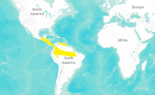
Figure 1: strawberry poison dart frog habitat region.

Strawberry poison dart frogs can easily be identified by their bright and vivid coloration. The entire body of the frog is a bright red-orange colour, save for it’s feet, and often times entire hind legs, which are a metallic gray-blue speckled with black. The frog is often referred to as the “blue-jeans” poison dart frog due to the colour of it’s hind legs. Relatively small in size, the Strawberry poison dart frog ranges from 17-24mm in length (Sandmeier 2001). The aposematic coloration of the frog is used as a warning mechanism to alert predators of it’s poisonous nature (Summer et. al. 2001). The coloration within the species is incredibly diverse, which is said to be due to sexual selection over time, however other research suggests their vivid coloration could have developed in correlation with their toxicity (Summer et. al. 2001). Despite their small size, their presence rarely goes unnoticed due to the characteristic “chirp” noise they often emit (Penner 2011).

medium.jpgFigure 2: strawberry poison dart frog common morph.

The term “poison dart frog” is derived from the frog’s toxin being used for coating the tips of indigenous tribes’ blow-darts. The toxins were generally extracted via one of two methods: the first was to simply rub the tip of the dart on the frog’s moist back, and the second is to heat the frog up to a temperature at which it would sweat and secrete the toxins, which would then be collected.

Poison_dart_frog-1.pngFigure 3: poison retrieval from poison dart frog.

The Strawberry poison dart frog is considered to be the most toxic in the Oophaga genus, but not the most poisonous among frog species. When agitated, the poison dart frog secretes a liquid containing alkaloids, a nitrogen based organic compound that causes blocking of the Na+ channels of the affected species’ nervous system, resulting in paralysis (Elder 2006). Certain similar dart frog species, such as the Golden dart frog (Phyllobates terribilis) are toxic enough to kill a 150lb. human with only 136 micrograms of it’s secretion (Elder 2006). Interestingly, the frogs themselves do not produce the toxins which they carry; these toxins are acquired through the small insects which they prey on, who have built up small toxin levels through the plants that they consume (Penner 2011).

Golden Orb weaver spider

The golden orb weaver spider is a unique, highly fascinating, venomous species of spider that can be found in many of the warmer regions of the world, from Southeast Asia to Australia, to the Americas all the way up to the United States (Weems 2004). Its name arises from the golden coloration of its silk, while it body colour ranges from silver to dark plum to orange, with brown, yellow-stripped legs (Australian Museum 2015).

The golden orb weaver spins one of the strongest and toughest webs in the world, and unlike most other spider species, their intricate webs are much like permanent homes, often lasting for multiple years (Ria Tan 2001). They use their webs, which are strung between trees in forests, to catch a variety of medium to large flying insects, such as wasps, moths, flies and butterflies (Weems 2004). Though they do not feed on them, their webs have even been known to entangle small birds! Birds also happen to be the main predator of this spider species. The male golden orb weaver, whose size is approximately 5mm, is significantly smaller than his female counterpart, who is usually 2-4 cm in length. Males have been known to take advantage of this difference in size by living on the female’s web, feeding off her catches without her even noticing. The male is even able to inseminate the female without her realizing, often while she is distracted by eating (Ria Tan 2001). Once fertilized, the female will dig a hole and bury her egg pouch underground. The golden orb weaver has a potent venom, which it injects into its prey through its chelicerae, or jaws, in order to paralyze it (Australian Museum 2015). Though the venom is a neurotoxin, it is not considered lethal to humans, and results only in redness, mild pain around the area of the bite, and a blister, which usually subsides after one full day.

12946937_1268914126456124_1.jpg
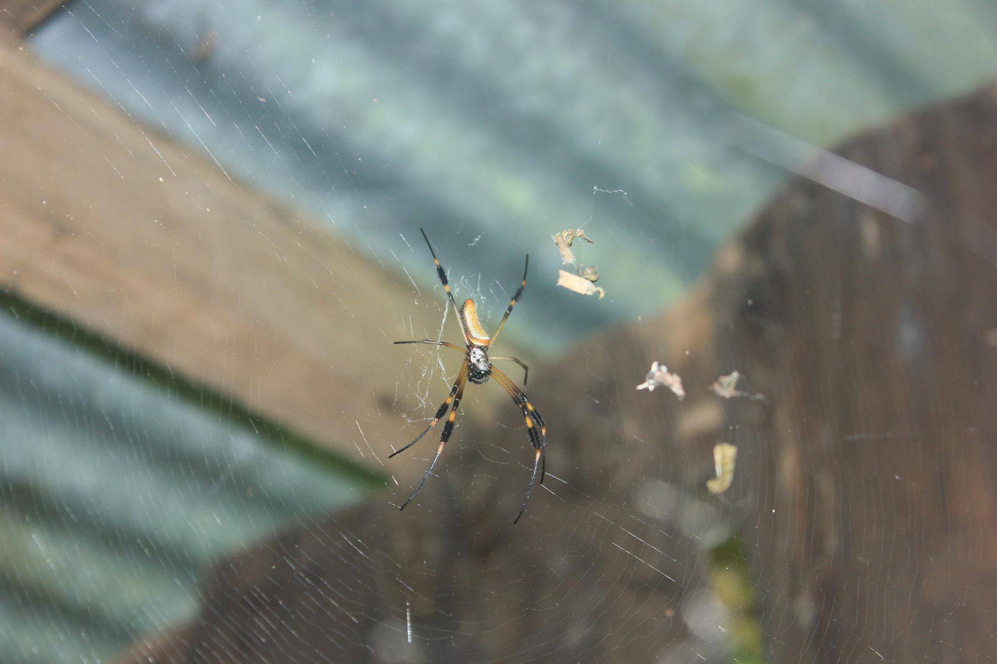
Figure 4: golden orb weaver spider

Fer-de-Lance snake

The Bothrops asper, or Fer-de-Lance, is a venomous snake native to tropical Central America (Fer-de-lance), favouring moist and wet environments over drier climates (Streiter), however, that is not to say they are not present in dry tropical regions as well. In Costa Rica, this species is most common in lowlands of both the Pacific and Caribbean coasts up to an elevation of 1300-m (Streiter).

Species of the Bothrops genus are recognizable by their noticeably broad, flat heads; the Fer-de-Lance are actually named so for their lance heads (Encyclopedia Britannica). They are also pit vipers, so they have heat sensitive moveable fangs that are positioned between each large eye and nostril (Brown 2011); these are used to accurately attack and target their warm-blooded prey (Encyclopedia Britannica). Their eyes are also recognizable by their large, vertical pupils (Streiter). Bothrops aspers also exhibit dimorphism; the females are noticeably larger than the males from birth, as well as have more ventral and subcaudals scales (Brown 2011). Both males and females have distinct coloration patterns consisting of pale cream-coloured diamond shaped patterned bands on their back and sides that complement their dark brown/grey scales (Streiter). They are usually 4-7 feet long, with the females being noticeably larger than males, both as juveniles and as infants, and can reach up to ten times the size of a male Fer-de-Lance (Brown 2011). Juvenile females also have brown-tail tips while juvenile males have yellow ones, which they completely shed after maturation (Brown 2011).

venomous_vs_non-venomous.jpgFigure 5: pit-viper vs. nonvenomous snake

12966757_10154154147504759_.jpg
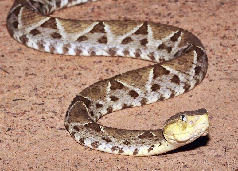
Figure 6: fer de lance snake

The Fer-de-Lance is considered the most dangerous snake in the Central America and is responsible for a majority of snake-induced deaths in humans (Strieter). Given their choice of diet, which ranges from small reptiles, invertebrates and arthropods as juveniles and slightly larger mammals as adults (Streiter), one could assume their natural defenses evolved to attack their prey more successfully, although this information has yet to be confirmed.

Manchineel tree

The Manchineel (Hippomane mancinella) is thought by many to be the worlds most dangerous tree. It is indigenous to Central America and the Caribbean (Sparman et. al. 2009). In central America the locals call it “Manzanita de la muerte” which means little apple of death (McLendon 2014). This name originates from the small apple shaped fruits produced by the tree. When ingested the fruits are reported to have a pleasantly sweet flavor. However, moments after ingestion tingling in the throat is felt, which quickly progresses into a burning sensation. Ingestion of the fruit ultimately leads to blistering of the mouth and throat and respiratory symptoms (Sparman et. al. 2009). The trees poison is not restricted to its fruit. Almost every part of the Manchineel including leaves, bark and sap are toxic. If skin comes in contact with Manchineel sap, bullous dermatitis and acute keratoconjunctivitis can occur (Sparman et. al. 2009). After brief exposure to the sap painful blisters can develop. The reason the Manchineel evolved such extreme toxic defenses is not yet confirmed. However, it is hypothesized that it may have something to do with how the tree reproduces. The Manchineel is located in costal areas and mangroves and uses the wind to disperse its seeds. Therefore, it is not necessary for animals to ingest the Manchineel fruits in order to spread seeds. Most animals are affected by the Manchineel’s potent toxins. However, certain species, like the iguana, are resistant.

12980864_1047206948688004_3.jpg
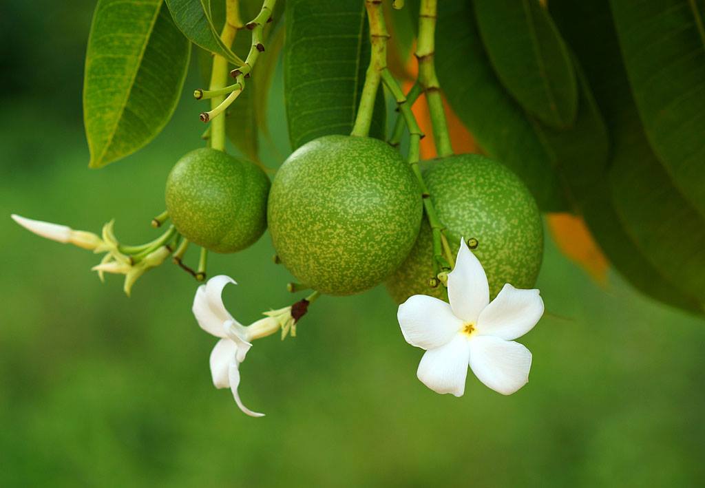
Figure 7: Manchineel tree’s poisonous apple

Fig 8 ToxicCE.jpgFigure 8: Manchineel tree

Costa Rica’s great biodiversity and subsequent high levels of competition and predation have lead to incredible biological adaptations. Many organisms have evolved to use toxins. Some, like the Poison Dart Frog and Manchineel tree, secret poison as a defense mechanism. Others, such as the Golden Orb Weaver spider and Fer-de-Lance, use poison for predation. There is a huge diversity in the toxins found in organisms. Some poisons are lethal, with the intention to kill the predator or prey. Others result in unpleasant reactions like blisters, meant to deter predators. Just as some organisms have evolved to use toxins to their advantage, others have developed immunity to these toxins.

References

Brown, Kelly. “Bothrops asper.” Animal Diversity Web. University of Michigan Museum of Zoology, 2011. Web. 01 Apr. 2016. http://animaldiversity.org/accounts/Bothrops_asper/

Elder, Jill. “Toxic Effects of Native Poison Dart Frogs (Dendrobatidae) in Costa Rica.” 2006 Costa   Rica Ecology Pre-Course Presentation Topic Reports. University of Miami of  Ohio, 18 May  2006. Web. 02 Apr. 2016. http://jrscience.wcp.miamioh.edu/fieldcourses06/PapersCostaRicaArticles/Toxiceffectsofnativepoiso.html

“Fer-de-lance.” Encyclopedia Britannica. Encyclopedia Britannica, n.d. Web. 02 Apr. 2016. http://www.britannica.com/animal/fer-de-lance-snake-genus

“Golden Orb Weaving Spider.” Australian Museum. Australian Museum, n.d. Web. 01 Apr.       2016. http://australianmuseum.net.au/golden-orb-weaving-spiders

“Golden Orb Web Spider.” Mangrove and Wetland Wildlife at Sungei Buloh Nature Park. Ria       Tan, 2011. Web. 01 Apr. 2016. http://www.naturia.per.sg/buloh/

IUCN “The IUCN Red List of Threatened Species. Version 2015-4”. iucnredlist.org.        Downloaded on 04 Apr. 2016. http://www.iucnredlist.org/details/55196/0

McLendon, Russell. “Why manchineel might be Earth’s most dangerous tree” Mother Nature Network. Mother Nature Network, 23 Oct. 2014. Web. 01 Apr. 2016. http://www.mnn.com/family/protection-safety/blogs/why-manchineel-might-be-earths-most-dangerous-tree

Penner, Austin. “Oophaga pumilio.” Animal Diversity Web. University of Michigan Museum of   Zoology, 2011. Web. 02 Apr. 2016. http://animaldiversity.org/accounts/Oophaga_pumilio/

“Pit viper.” Encyclopedia Britannica. Encyclopedia Britannica, n.d. Web. 02 Apr. 2016. http://www.britannica.com/animal/pit-viper

Sandmeier, Fran. “Oophaga pumilio: Strawberry Poison Frog”. Amphibiweb. UC Berkeley, 21   Mar. 2001. Web. 05 Apr. 2016. http://amphibiaweb.org/cgi/amphib_query?where-genus=Oophaga&where-species=pumilio

Sparman, John and L. Willis. “Manchineel poisoning bradyarrhythmia. A possible association.”   West Indian Medical Journal, Vol. 58 no. 1 (2009). carribean.scielo.org. Web. 03 Apr.2016. http://caribbean.scielo.org/scielo.php?script=sci_arttext&pid=S0043-31442009000100012

Streiter, Amy. “Fer-de-Lance.” Anywhere Costa Rica. Anywhere Costa Rica, n.d. Web. 02 Apr. 2016. http://www.anywherecostarica.com/flora-fauna/reptile/fer-de-lance

Summers, Kyle, and Mark E. Clough. “The Evolution of Coloration and Toxicity in the Poison     Frog Family (Dendrobatidae).” Proceedings of the National Academy of Sciences of the   United States of America. The National Academy of Sciences, 15 May 2001. Web. 05 Apr. 2016. http://www.pnas.org/content/98/11/6227.abstract

Weems, H.V. and G.B. Edwards. “Golden silk spider.” Featured Creatures: Entomology and       Nematology. University of Florida Institute of Food and Agricultural Studies, Aug. 2001.         Web. 01 Apr. 2016. http://entomology.ifas.ufl.edu/creatures/misc/golden_silk_spider.htm

Figures:

Figure 1: http://www.facts-about.info/poison-dart-frog/

Figure 2: http://animaldiversity.org/accounts/Oophaga_pumilio/

Figure 3: https://dixonapbio-taxonomywiki 2015.wikispaces.com/Poison+dart+frog+(chordate-+amphibian)

Figure 4: Lily Ragsdale, 2016.

Figure 5: http://asawright.org/venemous-snakes-of-trinidad-tobago/

Figure 6: http://ambergriscaye.com/photogallery/101229.html

Figure 7: http://www.planetdeadly.com/nature/most-poisonous-plants

Figure 8: http://fortlauderdaleforester.blogspot.ca/2013/01/tree-thursday_17.html

The Strangler Fig: The Ultimate Tree Hugger

by: Nicole Kitner, Ella Martin, Mikayla Salmon-Beitel, Carole-Anne Williams

In the depths of the Costa Rican rainforest, one can find many species of animals, insects, and plants. The interactions of these organisms vary, from the symbiotic relationship between hummingbirds and bromeliads, to the incredibly organized nest of leaf cutter ants, where they tend to their mutualistic relationship with fungi. Out of all the incredible wonders you may find on your journey through the rainforest, there is one species that seems to, quite literally, overtake the rest. The strangler fig trees are unlike most other trees in their environment. Their adaptations to utilize another tree’s resources, their unique relationship with wasps, and interactions with their ecosystem, make them an exceptionally fascinating species that have important impacts on their environment.

The strangler fig tree has certain adaptations to better suit its typical tropical rainforest environment. They are semi-deciduous, and only lose their foliage for short periods of time, usually during the dry season. They have a high drought tolerance, which is an adaptation that proves to be very important, particularly during the summer months when humidity and access to water is decreased (Brown). This tolerance enables survival, despite the less-than-optimal conditions during this period. The strangler fig’s foliage is entire, alternate, dark green, and somewhat leathery, and its leaf’s shape is elliptical to oblong, with cordate, acute, or obtuse bases, and a pointed apex (Brown). The pointed apex of the leaf is optimal in the tropical rainforest, allowing for easy runoff of water. Although the tree has no visible flowers, they are actually hidden within the hollow receptacle, which is called the fig. The pollination of these flowers is dependent on fig wasps, which have a symbiotic relationship with the tree (Prasertong).

The fig-fig wasp relationship is one of the key examples of coevolution and mutualistic symbiosis: each species of fig has evolved alongside a unique species of wasp that is small enough to crawl inside the fig through a tiny opening called the ostiole to lay its eggs inside, although they often may lose their wings or antennae in the process. In exchange, the wasp brings pollen into the fig, which is actually a cluster of seeds and flowers called an inflorescence (not a fruit), fertilizing the ovaries (Kline). Due to competition for a limited number of oviposition sites, the queen may visit three or four different figs to increase reproductive chances, but because she lost her wings they must be within close proximity to each other (Suleman, Nazia, et al.). Once her mission is complete, the queen then dies and is digested by the fig, providing it with nutrients. Once her eggs hatch, the males and females first mate with each other, then the females collect pollen to continue the process, while the males carve a path out of the fig to allow the females’ escape. The males are wingless and will spend their entire life within the fig (Kline).

Although it seems simple, several factors can arise to complicate this relationship. One is the possibility of uncooperative symbionts. In this case, one of the organisms (generally the wasp), fails to uphold its end of the deal (pollinating the fig), and becomes parasitic. If such a situation arises, the host fig tree can provide sanctions that limit the reproductive success of the cheating wasps. However, the tree is only able to target the entire fruit that are not performing, as opposed to individual wasps. Thus, pollen-free wasps are persistent in fig trees, albeit at low levels, as long as there are others within the same fig that can do their job for them (Jandér, et al.).

Another complication is specialized parasitic wasps that can lay their eggs within the fig from the outside using a long ovipositor, and that do not pollinate the fig in return. This leads to increased competition for oviposition sites, and possibly a lack of sites for the wasps that are pollinating. Something that reduces this effect is the presence of some ant species, which live on the fig tree and are able to move rapidly to catch flying insects, and actually patrol the fig to prey on the parasitic wasps. They have been shown to significantly reduce the wasps’ negative effect on the mutualism, and allow this relationship to continue (Schatz, Bertrand, et al.).

Strangler figs also participate in another form of symbiosis. This is their most notable, as well as arguably most interesting adaptation: it has the ability to steal another tree’s access to sunlight and nutrients by essentially killing the host tree. The strangler fig can grow from seeds, but in tropical rainforests, it has adapted to start as an epiphyte on another tree instead.

Picture1.jpg

At the epiphytic stage, the fig tree’s relationship with its host is seemingly innocent. Birds eat the fig’s fruit and drop its seeds between the leaf bases or in the nooks between branches of the host tree – usually palm and oak trees (Brown). The seedling grows there, finding nutrients in decaying leaves and soil, and growing towards the sunlight. This relationship is referred to as commensalism: the fig tree is benefitting from the host’s resources, while the host remains unaffected (Sack). As the epiphyte grows and sends its roots down along the trunk of the host, their relationship begins to change. When two root tendrils touch, they fuse together in a process called anastomose, eventually forming a massive woody mesh that completely encircles the trunk of the host. As the host continues to grow, the roots’ grip gets tighter, crushing its bark and constricting vital phloem and cambial layers (Armstrong). If the grip is tight enough, the host may no longer be able to transport nutrients throughout its system, dying of strangulation. Though the fig is not directly feeding on its host’s resources, it can be argued that their relationship at this point is parasitic, as the fig is harming the host tree in the hopes of eliminating competition for rain and sunlight (Sack). In most cases though, the host is sooner affected by the shading effects of the fully grown fig tree than by strangulation. This is especially true when the host is a palm tree, as palms lack an outer cambial and phloem layer (Armstrong).

The ficus family of trees, vines and shrubs are absolutely vital to many tropical ecosystems. One study recorded a total of 990 bird and 284 mammal species who eat figs as a part of their diet at one point during the year (Shanahan, Mike et al). These numbers include frugivores such as bats, monkeys, hornbills, toucans and parrots in addition to some invertebrates, that have been known to eat figs and aid in seed dispersal like the dung beetle, ants and, of course, fig wasps (Shanahan, Mike et al). Some of these animals rely completely on figs; they are called specialists (hornbills, some parrots and some toucans) and they would die out if figs disappeared. The majority of the animals that fall into the category of fig-eaters only do so at certain periods of the year when the main fruits of their diet are unavailable, or in times of food shortage. Despite the fact that figs are only eaten for a short period of time, in these cases they are vital: without them, these animals could starve (Kinnaird, M. ). In the food shortage of 1971 when only 50% of fruit trees in Barro Colorado Panama were producing fruit, the rates of starvation were better than they would have been due to fig trees in the area. In this case figs were a fallback food source for many species (Leigh, Egbert Giles).

Picture2.jpg

Ficus tree shading out its cabbage palm host in Fort Lauderdale, Florida        

There are several reasons why figs sustain so many animals in tropical rainforests. A big part is due to their fruiting patterns: most have very short periods between fruiting episodes, so there is fruit practically all year round. Also, they produce in the hundreds of thousands of figs that ripen at the same time, which increases the incentive of animals to feed there (Shanahan, Mike et al). The fruit itself comes in different colours and scents which caters to the variety of animals foraging for food. For example, the figs that ripen to be a deep red colour are easily spotted by birds whereas cauliflorous figs, that remain green but smell sweet, are scented at night by bats (Shanahan, Mike et al).

Picture3.jpgPicture4.jpg

Figs are also very easy for animals to eat. They do not have any protective coat ( no thorns or hard outer layer to dissuade hungry animals) and they are very nutritious. Although they do not contain high levels of carbohydrates or lipids like other fruits, they are much higher in protein, calcium and magnesium. Fig fruits contain 3.2 times more calcium than other tropical fruits, which is important for strong eggshells and increasing bone density (Kinnaird, M.). The protein often comes from the fig wasps inside the fruits, dead male wasps or eggs. Also, the sheer amount of available fruit per tree reduces competition between animals because there is more than enough for all (Shanahan, Mike et al). All in all, it is undeniable to say that if the ficus family disappears, many other animals would disappear with them.

The strangler fig is a unique species of tree that holds an important role in its tropical rainforest ecosystem. Over time, it has evolved a slew of adaptations that make it competitive to the many other species in its surroundings. Not only is the strangler fig physically well adapted to its environment, but it has also developed both mutualistic and parasitic relationships with other species in order to ensure its survival. Its fruit also serve as the main food source for many species of mammals and aves. The strangler fig is a perfect example of the complex adaptations necessary for survival in the tropical rainforest ecosystem. Due to its warm climate and fertile land, the tropical rainforest is a very productive environment, where plant species evolve twice as quickly as those in Antarctica (“Hotspots for Evolution”)! Because of this speedy rate of change, plant species have needed to develop complex interrelationships with other species in order to maintain a competitive advantage. Though strangulation may seem like a violent means by our standards, it is not in fact one of the most intense adaptations. The Acacia tree, for example, is covered with spikes housing colonies of poisonous ants that will not hesitate to attack the unsuspecting passerby. So unless you are Tarzan, we suggest being very careful when entering the rainforest jungle, as you never know what sneaky adaptation evolution will surprise you with next!

References:

Armstrong, Wayne P., “Stranglers and Banyans: Amazing Figs Of The Tropical Rain Forest.” Wayne’s Word. Palomar College, 1999. Web. 3 April 2016.

Brown, Stephen H. “Ficus Aurea.” (2011): n. pag. University of Florida IFAS Extension. University of Florida, 2011. Web. 2 Apr. 2016.

Brown, Stephen H. “The Prop and Buttress Roots of Banyan/Ficus Trees.” (2012): n. pag. University of Florida IFAS Extension. University of Florida. Web. 2 Apr. 2016.

Charlotte Jandér, K., et al. “Precision Of Host Sanctions In The Fig Tree-Fig Wasp Mutualism: Consequences For Uncooperative Symbionts.” Ecology Letters 15.12 (2012): 1362-1369. Academic Search Premier. Web. 15 Mar. 2016.

“Fig Wasps.” Figs Traditional Herbal Medicines for Modern Times The Genus Ficus (2010): n. pag. Web. 2 Apr. 2016.

“Hotspots for Evolution.” Understanding Evolution. University of California Museum of Paleontology, 2016. Web. 5 April 2016.

Kinnaird, M. The Ecology and Conservation of Asian Hornbills: Farmers of the Forest. Chicago: U of Chicago, 2008. Google Books. Web. 3 Apr. 2016

Kline, Katie. “The Story of the Fig and Its Wasp”. Ecological Society of America. ESA, 20 May 2011. Web. 15 Mar. 2016.

Laman, Timothy G (1995). “The ecology of strangler fig seedling establishment.” Selbyana, v. 16 issue 2, p. 223.

Leigh, Egbert Giles. Tropical Forest Ecology: A View from Barro Colorado Island. New York: Oxford UP, 1999. Google Books. Web. 3 Apr. 2016.

Prasertong, Anjali. “Strange Symbiosis: The Fig and the Wasp.” The Kitchn. N.p., 14 Sept. 2010. Web. 02 Apr. 2016.

Sack, Leo. “Growth and Ecology of Strangler Figs.” Tropical Ecosystems: Coral Reefs, Rainforests & A Potpourri of Weather, Earth Science & Other Good Things. Miami University, May 7 2014. Web. 3 April 2016.

Schatz, Bertrand, et al. “Complex Interactions On Fig Trees: Ants Capturing Parasitic Wasps As Possible Indirect Mutualists Of The Fig–Fig Wasp Interaction.” Oikos 113.2 (2006): 344-352. Academic Search Premier. Web. 15 Mar. 2016.

Shanahan, Mike, Samson So, Stephen G. Gompton, and Richard Gorlett. “Fig-eating by Vertebrate Frugivores: A Global Review.” Biological Reviews 76.4 (2001): 529-72. Web. 3 Apr. 2016.

Suleman, Nazia, et al. “Putting Your Eggs In Several Baskets: Oviposition In A Wasp That Walks Between Several Figs.” Entomologia Experimentalis Et Applicata 149.1 (2013): 85-93. Academic Search Premier. Web. 15 Mar. 2016.

Picture 1:  Ella Martin, 2016

Picture 2: Brown, Stephen H. “Ficus Aurea.” (2011): n. pag. University of Florida IFAS Extension. University of Florida, 2011. Web. 2 Apr. 2016.

Picture 3: Mulvihill, Robert. “Let’s Talk About Birds: Hornbills.” Post-Gazzette. Pittsburgh Post-Gazette, 13 Nov. 2013. Web. 08 Apr. 2016.(Link: http://www.post-gazette.com/news/science/2013/11/13/Let-s-Talk-About-Birds-Rhinoceros-hornbill/stories/201311130019)

Picture 4: Wilson, Dede. “Comparing Fresh Fig Varieties.” Bakepedia. Bakepedia, 24 Aug. 2013. Web. 08 Apr. 2016. (Link: http://www.bakepedia.com/tipsandtricks/comparing-fresh-fig-varieties/)


Baird’s Tapir in Costa Rica: In Danger of Extinction

By Jessica Di Bartolomeo, Michelle Rijski and James Theodore

IMG_20160112_091210Figure 1: Mural of Baird’s Tapir at Rio Celeste, Bijagua.

Baird’s Tapir are the largest mammals in Central America. They were declared in danger of extinction by the International Union for Conservation of Nature and Natural Resources (IUCN) in 2002. As one of the countries home to Baird’s Tapir populations, Costa Rica must use effective conservation strategies to ensure extinction does not occur.

Overview

Many threats such as deforestation have a great impact on Baird’s Tapir’s populations. Smaller factors such as poaching, cattle farming and agriculture also play a role in the decline. Due to their low and slow reproductive rate, they cannot rapidly regenerate the lives of their population. To prevent further loss, strategies must be put in place in order to protect the Baird’s Tapir’s population. Limiting deforestation, enforcing rules against hunting, reducing cattle farming and agricultural farms where Baird Tapirs are found are all possibilities to help their population regenerate.

General Information

Physical description and classification

Tapirus Bairdii, commonly referred to as Baird’s Tapir, belongs to the family Tapiridae and is one of four remaining Tapir species, all of which are threatened or endangered (Brooks, Bodmer and Matola; Terwilliger 1978). It is the least subjected member of its family to research (Terwilliger 1978). Baird’s Tapir is the largest native mammal in Central America (Tobler 2002; Foerster and Vaughan 2002), with an average weight of 150-320 Kg, a length of 180 – 250 cm, and a height of 73-120 cm. Juveniles possess a red-brown coloration with light coloured stripes and spots for camouflage. As they mature, their thick bristly fur becomes a fully dark brown or grey. Tapirs have a long snout, stocky bodies and strong legs adapted for moving quickly in dense vegetation (EDGE 2010). A distinguishing characteristic is their long snout, called a proboscis which extends past their chin and resembles a shortened elephant trunk. It is used to forage through vegetation, picking up eatable plants and bringing them to their mouths (EDGE 2010).

Photo_2016-04-06_4_21_41_PM

Figure 2: Tapirus bairdii. Tapirs.org

Habitat, distribution and population

Baird’s Tapir is found from southeastern Mexico through to northwestern Columbia and the Andes of Ecuador (Tobler 2002), ranging from sea level to 3600 meters in altitude, and has been wiped out completely in El Salvador (Cuaron, Matola and Rubio-Tolgler 1997).

It is very difficult to directly observe Tapirs due to their small numbers, preference for dense vegetation and their brown hide that enables them to blend in with their surroundings (Fragoso 1987). When studying Tapirs, researchers often use indirect methods such as tracing tracks and feces. This has shown the Tapir’s habitat preference to be dense forests with bodies of water, such as tropical sub-deciduous forests and cloud forests, as opposed to pine forests and grasslands, which are drier and more exposed. Foerster and Vaughan’s study (2002) indicated a preference for secondary forest, likely because these habitats consist of a dense understory, therefore provides the Tapir with a multitude plants at accessible heights for consumption. Secondary forests would also have fewer forest fires and minimal human disturbance (Naranjo 2009). Studies conducted in Mexico and Costa Rica found that undisturbed areas had twice as many Tapirs. They tend to keep away from hiking trails as well as villages, such as Villa Mills in Costa Rica, where they have been hunted in the past. (Naranjo 2006). These studies also showed that no Tapirs are found near agricultural lands, whose formation by deforestation destroys the Tapirs’ habitat.

Photo_2016-04-06_5_46_14_PM

Figure 3: Map of Tapir Habitat. Ultimate Ungulate

Diet

Tapirs eat the stems, leaves, and fruits from over 100 species of plants, from various families including Poacae, Fagaceae, Solanaceae, and Rubiaceae. Studies have reported that they “forage in a zig zag pattern” (Terwilliger 1978; Cuaron, Matola and Rubio-Tolgler 1997). Although Tapirs have been found to prefer certain plant species, their broad diet allows them to live in a variety of habitats and at a wide range of elevations (Naranjo 2006). Tapirs are considered to be seed predators, because many seeds they consume are either destroyed through chewing and digestion or dispersed through expulsion. Tapirs also play an important role in seed dispersion by either spitting out the seeds from fruit they eat, or by defecating them. Baird’s Tapir is important for the maintenance of swamp ecosystems in Costa Rica and Nicaragua because of their effective ability to disperse seeds. In fact, Tapirs can disperse hundreds to thousands of seeds at a time, which have been found sprouting from their feces, often many kilometres away from the parent plants. Furthermore, Tapirs tend to defecate in streams, often further promoting plant growth (Brooks, Bodmer and Matola).

Mating

Tapirs have a 13-month gestation period and produce a single offspring, called a calf, which remains with its mother for the first two years of its life, until it reaches sexual maturity. For the first 10 days or so the calf remains hidden while its mother retrieves food; it then starts following its mother around. Females can have babies until they are approximately 10 years of age, and at most one baby every 14 months. Their low rate of reproduction is a contributing factor in population decline (Brooks, Bodmer and Matola).

Photo_2016-04-06_5_38_47_PMFigure 4: Tapir calf. Prague zoo

Behaviour

Tapirs are very shy and generally solitary but have been found to come together to feed and drink, where they communicate through vocalizations, whistles, and nose touching (Brooks, Bodmer and Matola; Terwilliger 2002). To locate one another, they usually use their sense of smell, or call out with a high-pitched whistle (Brooks, Bodmer and Matola). Although Tapirs are neither distinctly nocturnal nor diurnal, they tend to be most active at night (EDGE 2010). One study found that Tapirs were active 20.2% of the time during the day and 80.4% at night (IUCN 2002). It has been suggested that the Tapir’s behaviour is an adaptation to avoid the heat, as the large mammal would not be efficient at eliminating heat to return to its homeostatic temperature. Furthermore the Baird’s Tapir’s nocturnal activity would allow them to steer clear from humans (Foerster and Vaughan 2002). Tapirs are also highly sensitive to and fearful of noise, resulting in an avoidance of areas with greater human activity (Terwilliger 1978).

Threats

The International Union for Conservation of Nature and Natural Resources (IUCN) categorizes Baird’s Tapir as endangered because of its significant decrease in numbers (IUCN 2002). Its declining population is primarily due to habitat destruction but hunting contributes to the loss (Cuaron, Matola and Rubio-Tolgler 1997; Foerster and Vaughan 2002). Estimates have shown there to be less than 5,500 Baird’s Tapirs left in the wild, with the majority in Mexico (over 1500), Guatemala, Costa Rica and Panama (each with less than 1000) (IUCN 2002). Because of deforestation and habitat fragmentation, Tapirs now mostly live in protected areas and regions not easily accessible to humans. It is not certain whether these areas are adequately large enough to sustain Tapir populations long term (Naranjo 2009). Tapirs’ low reproductive rates make them especially vulnerable to extinction (Cuaron, Matola and Rubio-Tolgler 1997). Decreases in population size slowly recover; therefore, changes in their environment can cause great damage to the species’ survival.

Main threat: Effect of Deforestation

Central America today is home to 70% less forested regions than 40 years ago. Since Baird’s Tapir lives in these forests, much of its habitat has been lost. As forests become more fragmented, the general trend for Tapir populations is to decline. Tapir habitat, including rivers and areas with dense vegetation, are increasingly being transformed into land suitable for logging, farming and agriculture. The remaining forest fragments thus contain a smaller, isolated Tapir population. Many of these forest fragments in Mexico have shown a sharp rise in other species like collared peccaries and white-tailed deer, which Tapirs must compete with for food (Naranjo 2009).

Other threats: Effects of Cattle Farms and Agriculture

Cattle farms are potential source of disease which can harm Perissodactyla populations such as Tapirus Bairdii. The fences put in place for these farms also inhibit Tapirs’ movement (Cuaron, Matola and Rubio-Tolgler 1997). Furthermore, pesticide and fertilizer runoff pollute drinking water, and soil erosion on hills and steep slopes can be deadly (Naranjo 2009). Tapirs must also compete for food with free ranging farm animals like cattle and horses.

Photo_2016-04-06_4_24_07_PMFigure 5: Baird’s Tapir in Water. Los Angeles Zoo.

Conservation

Baird’s Tapir in Costa Rica are likely found in the following areas: Arenal, Cordillera Volcanica Central, La Amistad, Corcovado, Osa, Guanacaste-Santa Rosa, and Llanuras de Tortuguero (Cuaron, Matola and Rubio-Tolgler 1997; Tobler 2002). More research is needed to map out Tapir presence throughout Costa Rica in order to determine with more precision the locations where they have survived and where they have been eliminated (Cuaron, Matola and Rubio-Tolgler 1997). Current studies are also needed for more accurate quantifications of Tapir population in Costa Rica. The most recent study to quantify the number of Baird’s Tapir in Costa Rica, which estimated the population reached no more than 1000, took place in 1990 (Vaughan 1990).

Preservation

A main goal for protection of Baird’s Tapir is habitat preservation, which includes reducing forest fragmentation. Since Tapirs’ habitat usually approaches water sources, the quality of these bodies is vital to conserve as well. Monitoring and reduction of harmful chemicals in agriculture should be an important part of conservation efforts (Foerster and Vaughan 2002).

Hunting

In 1992, the Costa Rican government passed a conservation law which protects Baird’s Tapir and other species from hunting. Current data is needed to verify the upholding of hunting-prohibition laws.

Because of their naturally low density and low birth rate, it is not believed possible for Tapirs to be hunted sustainably. Communities that hunt Tapirs for meat must switch to species with higher densities and birth rates, such as white-tailed deer, peccaries, and armadillos (Naranjo 2009).

In Mexico, Tapir populations are continuing to become isolated from each other, despite an increase in protected regions. This is likely because laws concerning unsustainable practices such as overhunting and crop burning are not properly imposed. Policy-makers are inconsistent in the laws they implement to protect endangered areas and frequently grant subsidies for conventional farming and cattle grazing. Conventional farming should also be discouraged and replaced by sustainable cattle ranches, organic agriculture and agroforestry (Naranjo 2009).

Environmental Education

If rural communities were to get involved in conservation efforts, the Tapir’s status could significantly improve. Jobs like ecotourism, Tapir observation, and feces and track counting should be introduced into these communities. Additionally, communities could host captive breeding areas for Tapirs, which would attract tourists and promote conservation awareness. Environmental education should be encouraged by governments to be taught in schools from elementary through to the end of high school and beyond. Schools and other institution should offer wildlife management courses to rural residents as well as students in larger cities (Naranjo 2009).

References

  1. Brooks, D. M., Bodmer, R. E. and Matola, S. Tapirs. [Online]. HTTPS://portals.iucn.org [2016, Jan 29]
  2. Cuaron, Alfredo D., Sharon Matola and Heidi Rubio-Tolgler (1997).Status and Action Plan of Baird’s Tapir (Tapirus bairdii) [16pp]. IUCN Tapirs Status Survey and Conservation Action Plan. [Online]. Available: https://portals.iucn.org/library/efiles/documents/1997-053.pdf [2016, Feb. 4].
  3. EDGE (12 November 2010). Baird’s Tapir. [12 pp.] [Online]. Available: http://www.edgeofexistence.org/mammals/species_info.php?id=34 [2016, Jan. 29]
  4. Foerster, Charles R and Vaughan, Christopher (2002, September). Home Range, Habitat Use, and Activity of Baird’s Tapir in Costa Rica. [14pp] Biotropica [Online serial], 34(3). Available: http://www.jstor.org/stable/4132941 [2016, Feb. 11].
  5. Fragoso, Jose Manuel V. (1987). The Habitat Preferences and Social Structure of Tapirs [101pp]. Master’s Thesis; University of Toronto. Available: http://web.stanford.edu/group/fragoso/docs/Fragoso%20JMV%201987%20MS%20thesis%202.pdf [2016, Jan. 30].
  6. IUCN (2002). Tapirus bairdii. [Online]. Available: http://www.iucnredlist.org/details/21471/0 [2016, Feb. 3].
  7. Los Angeles Zoo. Tapir, Baird’s. Los Angeles Zoo and Botanical Gardens. [Online]. Available: http://www.lazoo.org/animals/mammals/tapir-bairds/ [2016, April 6].
  8. Naranjo, Eduardo J. (2009, May 25). Ecology and Conservation of Baird’s Tapir in Mexico [19pp]. Tropical Conservation Science [Online serial], 2(2). Available: http://tropicalconservationscience.mongabay.com/content/v2/09-05-25_140-158_naranjo.pdf [2016, Jan. 30].
  9. Naranjo, E.J., M.W. Tobler and I Lira-Torres. (2006). Habitat Preference, Feeding Habits and Conservation of Baird’s Tapir in Neotropical Montane Oak Forests. In Ecology and conservation of neotropical montane oak forest. [Online]. Available: https://www.researchgate.net/publication/225961764_Habitat_Preference_Feeding_Habits_and_Conservation_of_Baird’s_Tapir_in_Neotropical_Montane_Oak_Forests [2016, Jan. 30].
  10. Tobler, Mathias W. (2002, September). Habitat Use and Diet of Baird’s Tapir (Tapirus bairdii) in a Montane Cloud Forest of the Cordillera de Talamanca, Costa Rica. [7pp] Biotropica [Online serial], 34(3). Available: http://www.jstor.org/stable/4132948. [2016, Feb. 11].
  11. Prague Zoo. Prague Zoo Celebrates Newest Tapir Calf. [Online]. Available: http://www.zooborns.com/zooborns/2015/05/prague-zoo-celebrates-newest-tapir-calf.html [2016, April 5].
  12. Tapirs.org. The World’s Tapirs–The Baird’s Tapir (Tapirus bairdii). Tapir Specialist Group. [Online]. Available: http://www.tapirs.org/tapirs/bairds.html [2016, April 5].
  13. Terwilliger, Valery J. (September 1978). Natural History of Baird’s Tapir on Barro Colorado Island, Panama Canal Zone. [9pp] Biotropica [Online serial], 10(3). Available: http://www.jstor.org/stable/2387906 [2016, Feb. 11].
  14. Ultimate Ungulate. Baird’s tapir, Central American tapir. An Ultimate Ungulate Fact Sheet. [Online]. Available: http://www.ultimateungulate.com/Perissodactyla/Tapirus_bairdii.html [2016, April 5].

Figures:

Figure 1: J. Di Bartolomeo, 2016.

Figure 2: http://www.tapirs.org/tapirs/bairds.html

Figure 3: http://www.ultimateungulate.com/Perissodactyla/Tapirus_bairdii.html

Figure 4: http://www.zooborns.com/zooborns/2015/05/prague-zoo-celebrates-newest-tapir-calf.html

Figure 5:http://www.lazoo.org/animals/mammals/tapir-bairds/

Shedding a Light on Bioluminescence

by Julia Cohen, Michelle Gervais, Alexandra Moisan.

Bioluminescence is an emission of light caused by an oxidation reaction that occurs in living organisms. Many diverse microbes and marine animals are able to produce their own light, predominantly those concentrated in the ocean. This light production can be used as a means of communication among a species. The compounds involved in the chemical reaction are luciferin and either luciferase or photoprotein (1). Luciferin produces the light and the luciferase or photoprotein acts as the catalyzing protein of the oxidation reaction (6). The light given off is referred to as cold light as it is 100% light energy. This means that none of it’s energy is transformed into heat (5).

Adaptations

Feeding

Many deep sea fish will use bioluminescence as a lure in order to attract prey. Anglerfish, for example, grow a long appendage that hangs in front of them is used to bait smaller fish into swimming within striking distance. Other fish also use bioluminescence to attract their prey; the cookie cutter shark’s entire body is lit up except for a small patch on it’s stomach that resembles the shadow of a smaller fish. This small shadow-like patch lures in larger predatory fish, who upon attempting to attack what they believe to be a smaller fish, are met with the cookiecutter shark that preys on them (2).


Attraction

Many marine animals use bioluminescence to attract mates. Ostracods, or crustaceans that can be found in the western Caribbean sea, use a complex combination of lights to attract their mates. There are over a dozen different species of Ostracods living within similar habitats. In all of these crustacean species only the males produce bioluminescent light and yet their mating displays are all very different (4).

A non aquatic example of bioluminescent attraction occurs in fireflies. These insects use their bioluminescence to attract a mate and, similarly to the crustaceans each species has its own distinct lighting pattern. The differing patterns allow the female to be drawn to a male of their own species (5).

Protection

Bioluminescence is used by some animals to scare off predators or to distract them long enough to escape. Swima Bombiviridis, also known as the green bomb worm, releases a flash of bright green bioluminescent light when threatened. The same tactic is used by certain squid that produce bioluminescent liquid instead of ink. They release this liquid to distract predators while they escape (2).

Evolution

It is estimated that bioluminescence has evolved at least 40 times independently, but most likely 50 times among extant organisms. Non Symbiotic luminous organisms possess the gene for either luciferase or photoprotein, which are the compounds required to produce bioluminescence. It is believed that for bacterial symbionts, the trait may have evolved only once, but each marine animal lineage that uses those microbes has had to develop specialized organs to host the light and maintain the bacterial culture (10). A type of luciferin known as coelenterazine occurs in many marine bioluminescent groups. It previously had antioxidant properties that helped defend aquatic species from oxidative stress. Oxidative stress is a chemical imbalance that occurs in a body and is cause by free radicals. At some point there was a shift from its antioxidative function to the light emitting function that exists today. This is most likely due to the fact that the need for antioxidative defence mechanisms decreased when marine animals began inhabiting deeper waters. In these waters, the animals were less exposed to oxidative stress because there is less light and oxygen available. Within the organisms, there was a reduction in metabolic activity as they increased their depth in the sea. As a result, through evolution, these organisms developed mechanisms to create the bioluminescence produced by coelenterazine that can be seen today (9).

Dinoflagellates of Costa Rica

Screen Shot 2016-04-10 at 10.24.30 AM.png

In Costa Rica we observed bioluminescent dinoflagellates, which are a type of plankton. Ninety percent of all dinoflagellates are marine plankton. Dinoflagellates are approximately 0.04 mm in size, making them invisible to the naked eye, and are present in large agglomerations in the epipelagic zone of the ocean which is located up at the surface (7). Dinoflagellates are motile, they move by means of two flagella, a protein and microtubule strands which propel it through the water. They have an internal skeleton made of cellulose like plates (8).

Dinoflagellate Bioluminescence Chemistry

These tiny marine organism produce light through a luciferin-luciferase reaction. The luciferase found in dinoflagellates is related to the green chemical chlorophyll found in plants (1). Although the chemistry of the dinoflagellates luminescence is not completely understood, it is most likely caused by a drop in pH due to an influx of protons within its cell. It is an extremely fast cellular process and the dinoflagellate can produce a flash of light lasting up to 100 ms. However, once all its luciferin has been oxidized, it must wait until the following day for its chemicals to recharge to be able to flash again. (8) The cell alternates between photosynthesis and luminescence on a 24 hour cycle.

Ecological Roles and Relationships

Dinoflagellates use their luminescence for many different ecological functions. These protists can be autotrophic or heterotrophic. Photosynthetic dinoflagellates are important primary producers in coastal waters; some are symbiotic living in their hosting coral cells. Seen as they are small photosynthetic organisms, they are at the bottom of the food chain. Because of this, many aquatic creatures prey on them. Therefore, they primarily use their bioluminescence as a defence mechanism. By emitting light when stimulated by movement they can scare away smaller predators. They also act as a signal to larger predators. When they light up they draw the larger predators in, signaling the presence of a smaller predator they can prey on. This indirectly protects the dinoflagellates, creating a mutualistic relationship with the larger coastal predators (7).

References

  1. “Bioluminescence.” National Geographic Education. National Geographic, 13 June 2013. Web. 25 Mar. 2016. <http://education.nationalgeographic.org/encyclopedia/bioluminescence/&gt;.
  1. ”Adaptations for Bioluminescence – Bioluminescence.” Adaptations for Bioluminescence. Smithsonian National Museum of Natural History, n.d. Web. 25 Mar. 2016. <http://www.biology-online.org/articles/bioluminescence/adaptations-bioluminescence.html&gt;.
  1. ”Dinoflagellate Bioluminescence.” Latz Laboratory. Scripps Institute of Oceanography, 09 June 2014. Web. 25 Mar. 2016. <https://scripps.ucsd.edu/labs/mlatz/bioluminescence/dinoflagellates-and-red-tides/dinoflagellate-bioluminescence/&gt;.
  1. Rivers, Trevor J., and James G. Morin. “Complex Sexual Courtship Displays by Luminescent Male Marine Ostracods | Journal of Experimental Biology.” Complex Sexual Courtship Displays by Luminescent Male Marine Ostracods | Journal of Experimental Biology. Journal of Experimental Biology, n.d. Web. 27 Mar. 2016. <http://jeb.biologists.org/content/211/14/2252.long&gt;.
  1. United States. National Park Service. “Synchronous Fireflies.” National Parks Service. U.S. Department of the Interior, n.d. Web. 27 Mar. 2016. <https://www.nps.gov/grsm/learn/nature/fireflies.htm&gt;.
  1. Nealson, K. H., and John W. Hastings. “Bacterial bioluminescence: its control and ecological significance.” Microbiological reviews 43.4 (1979): 496.
  1. Wilson, Thérèse. Bioluminescence. Harvard University Press, 2013.
  1. ”Dinoflagellate Bioluminescence.” Latz Laboratory. Scripps Institution of Oceanography. UC San Diego, 09 June 2014. Web. 05 Apr. 2016.
  1. Others, J.-F. Rees. “THE ORIGINS OF MARINE BIOLUMINESCENCE: TURNING OXYGEN DEFENCE MECHANISMS INTO DEEP-SEA COMMUNICATION TOOLS.” The Journal of Experimental Biology 201, 1211–1221 (1998): 1211. Web. 5 Apr. 2016.
  1. Haddock, Steven H.D, Mark A. Moline, and James F. Case. “Bioluminescence in the Sea.” Marine Science 2 (2009): 443-49. Web. 5 Apr. 2016.

Picture 1: http://inhabitat.com/5-bioluminescent-species-that-light-up-the-world/

Picture 2: http://knowledgebase.lookseek.com/Angler-Fish-teleost-order-Lophiiformes.html

Picture 3: http://beneficialbugs.org/bugs/Firefly/boreal_firefly.htm

Picture 4: http://matadornetwork.com/trips/15-places-to-see-bioluminescence-pics/

Picture 5: http://visualsunlimited.photoshelter.com/image/I0000duZGSLe8aoU